Bacteria Cell Structure
They are as unrelated to human beings as living things can be, but bacteria are essential to human life and life on planet Earth. Although they are notorious for their role in causing human diseases, from tooth decay to the Black Plague, there are beneficial species that are essential to good health.
For example, one species that lives symbiotically in the large intestine manufactures vitamin K, an essential blood clotting factor. Other species are beneficial indirectly. Bacteria give yogurt its tangy flavor and sourdough bread its sour taste. They make it possible for ruminant animals (cows, sheep, goats) to digest plant cellulose and for some plants, (soybean, peas, alfalfa) to convert nitrogen to a more usable form.
Bacteria are prokaryotes, lacking well-defined nuclei and membrane-bound organelles, and with chromosomes composed of a single closed DNA circle. They come in many shapes and sizes, from minute spheres, cylinders and spiral threads, to flagellated rods, and filamentous chains. They are found practically everywhere on Earth and live in some of the most unusual and seemingly inhospitable places.
Evidence shows that bacteria were in existence as long as 3.5 billion years ago, making them one of the oldest living organisms on the Earth.
Even older than the bacteria are the archeans (also called archaebacteria) tiny prokaryotic organisms that live only in extreme environments: boiling water, super-salty pools, sulfur-spewing volcanic vents, acidic water, and deep in the Antarctic ice. Many scientists now believe that the archaea and bacteria developed separately from a common ancestor nearly four billion years ago. Millions of years later, the ancestors of today's eukaryotes split off from the archaea. Despite the superficial resemblance to bacteria, biochemically and genetically, the archea are as different from bacteria as bacteria are from humans.
In the late 1600s, Antoni van Leeuwenhoek became the first to study bacteria under the microscope. During the nineteenth century, the French scientist Louis Pasteur and the German physician Robert Koch demonstrated the role of bacteria as pathogens (causing disease).
The twentieth century saw numerous advances in bacteriology, indicating their diversity, ancient lineage, and general importance. Most notably, a number of scientists around the world made contributions to the field of microbial ecology, showing that bacteria were essential to food webs and for the overall health of the Earth's ecosystems. The discovery that some bacteria produced compounds lethal to other bacteria led to the development of antibiotics, which revolutionized the field of medicine.
There are two different ways of grouping bacteria. They can be divided into three types based on their response to gaseous oxygen. Aerobic bacteria require oxygen for their health and existence and will die without it. Anerobic bacteria can't tolerate gaseous oxygen at all and die when exposed to it. Facultative aneraobes prefer oxygen, but can live without it.
The second way of grouping them is by how they obtain their energy. Bacteria that have to consume and break down complex organic compounds are heterotrophs. This includes species that are found in decaying material as well as those that utilize fermentation or respiration. Bacteria that create their own energy, fueled by light or through chemical reactions, are autotrophs.
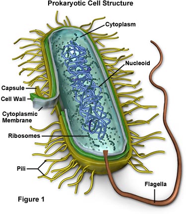
- Capsule - Some species of bacteria have a third protective covering, a capsule made up of polysaccharides (complex carbohydrates). Capsules play a number of roles, but the most important are to keep the bacterium from drying out and to protect it from phagocytosis (engulfing) by larger microorganisms. The capsule is a major virulence factor in the major disease-causing bacteria, such as Escherichia coli and Streptococcus pneumoniae. Nonencapsulated mutants of these organisms are avirulent, i.e. they don't cause disease.
- Cell Envelope - The cell envelope is made up of two to three layers: the interior cytoplasmic membrane, the cell wall, and -- in some species of bacteria -- an outer capsule.


- Cell Wall - Each bacterium is enclosed by a rigid cell wall composed of peptidoglycan, a protein-sugar (polysaccharide) molecule. The wall gives the cell its shape and surrounds the cytoplasmic membrane, protecting it from the environment. It also helps to anchor appendages like the pili and flagella, which originate in the cytoplasm membrane and protrude through the wall to the outside. The strength of the wall is responsible for keeping the cell from bursting when there are large differences in osmotic pressure between the cytoplasm and the environment.

Cell wall composition varies widely amongst bacteria and is one of the most important factors in bacterial species analysis and differentiation. For example, a relatively thick, meshlike structure that makes it possible to distinguish two basic types of bacteria. A technique devised by Danish physician Hans Christian Gram in 1884, uses a staining and washing technique to differentiate between the two forms. When exposed to a gram stain, gram-positive bacteria retain the purple color of the stain because the structure of their cell walls traps the dye. In gram-negative bacteria, the cell wall is thin and releases the dye readily when washed with an alcohol or acetone solution.
- Cytoplasm - The cytoplasm, or protoplasm, of bacterial cells is where the functions for cell growth, metabolism, and replication are carried out. It is a gel-like matrix composed of water, enzymes, nutrients, wastes, and gases and contains cell structures such as ribosomes, a chromosome, and plasmids. The cell envelope encases the cytoplasm and all its components. Unlike the eukaryotic (true) cells, bacteria do not have a membrane enclosed nucleus. The chromosome, a single, continuous strand of DNA, is localized, but not contained, in a region of the cell called the nucleoid. All the other cellular components are scattered throughout the cytoplasm.

- Flagella - Flagella (singular, flagellum) are hairlike structures that provide a means of locomotion for those bacteria that have them. They can be found at either or both ends of a bacterium or all over its surface. The flagella beat in a propeller-like motion to help the bacterium move toward nutrients; away from toxic chemicals; or, in the case of the photosynthetic cyanobacteria; toward the light.

- Nucleoid - The nucleoid is a region of cytoplasm where the chromosomal DNA is located. It is not a membrane bound nucleus, but simply an area of the cytoplasm where the strands of DNA are found. Most bacteria have a single, circular chromosome that is responsible for replication, although a few species do have two or more. Smaller circular auxiliary DNA strands, called plasmids, are also found in the cytoplasm.

- Pili - Many species of bacteria have pili (singular, pilus), small hairlike projections emerging from the outside cell surface. These outgrowths assist the bacteria in attaching to other cells and surfaces, such as teeth, intestines, and rocks. Without pili, many disease-causing bacteria lose their ability to infect because they're unable to attach to host tissue. Specialized pili are used for conjugation, during which two bacteria exchange fragments of plasmid DNA.


- Ribosomes - Ribosomes are microscopic "factories" found in all cells, including bacteria. They translate the genetic code from the molecular language of nucleic acid to that of amino acids—the building blocks of proteins. Proteins are the molecules that perform all the functions of cells and living organisms. Bacterial ribosomes are similar to those of eukaryotes, but are smaller and have a slightly different composition and molecular structure.


No comments:
Post a Comment